Welcome to the Amira-Avizo Software Use Case Gallery
Below you will find a collection of use cases of our 3D data visualization and analysis software. These use cases include scientific publications, articles, papers, posters, presentations or even videos that show how Amira-Avizo Software is used to address various scientific and industrial research topics.
Use the Domain selector to filter by main application area, and use the Search box to enter keywords related to specific topics you are interested in.
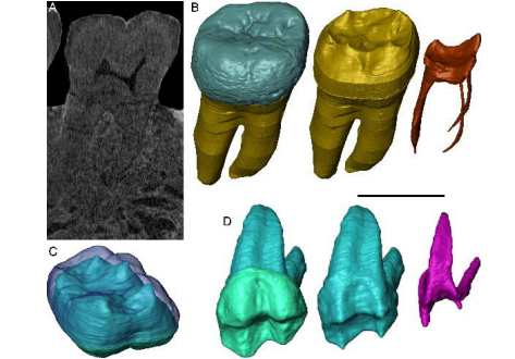
Exploring hominin and non-hominin primate dental fossil remains with neutron microtomography
Fossil dental remains are an archive of unique information for paleobiological studies. Computed microtomography based on Xray microfocus sources (X-µCT) and Synchrotron Radiation (SR-µCT) allow subtle quantification at the micron and sub-micron scale of the meso- and microstructural signature imprinted in the mineralized tissues, such as enamel and dentine, through highresolution “virtual histology”. Nonetheless, depending on the degree of alterations undergone during fossiliza... Read more
Clément Zanolli, Laboratory AMIS, UMR 5288, University of Toulouse III - Paul Sabatier, France, and al.
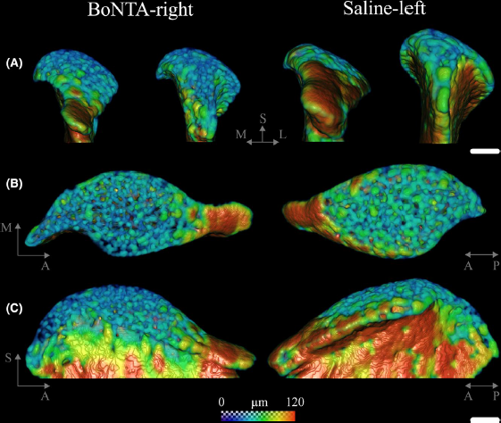
Masseter muscle function influences mandibular bone homeostasis. As previously reported, bone resorption markers increased in the mouse mandibular condyle two days after masseter paralysis induced with botulinum toxin type A (BoNTA), followed by local bone loss.
This study aimed to evaluate the bone quality of both the mandibular condyle and alveolar process in the mandible of adult mice during the early stage of a BoNTA‐induced masseter muscle atrophy, using a combined 3D histomorpho... Read more
Julián Balanta‐Melo, María Angélica Torres‐Quintana, Maximilian Bemmann, Carolina Vega, Constanza González, Kornelius Kupczik, Viviana Toro‐Ibacache, Sonja Buvinic
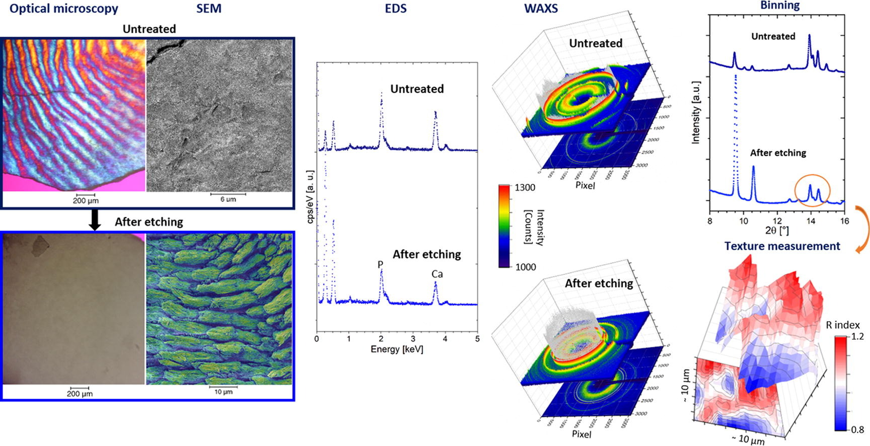
Enamel caries is a highly prevalent worldwide disease that involves the demineralisation of the outer tooth structure. In this study, we report the analysis of artificially demineralised human enamel sections (‘slices’) etched using lactic acid (2% v/v) in comparison with healthy enamel using correlative techniques of optical and electron microscopy, as well as scanning diffraction. Demineralisation of the enamel was characterised at the micron to sub-micron scale. The structure of the he... Read more
Cyril Besnard, Robert A. Harper, Thomas E. J. Moxham, Jonathan D. James, Malte Storm, Enrico Salvati, Gabriel Landini, Richard M. Shelton, Alexander M.Korsunsky
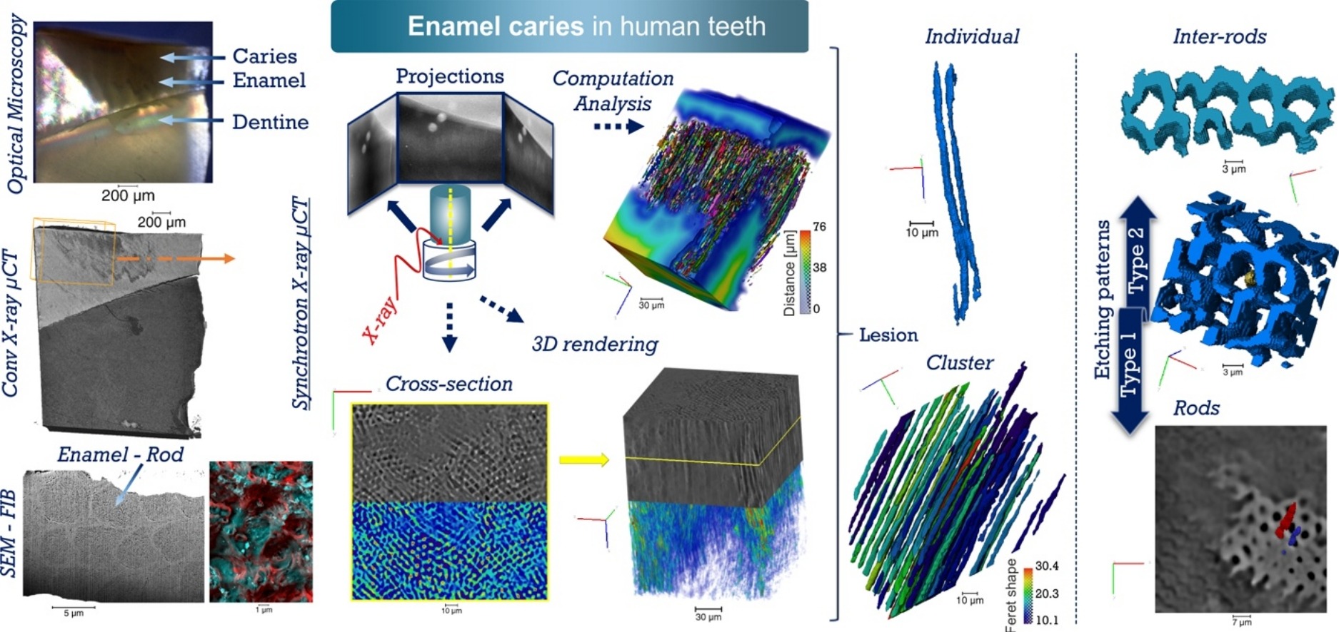
Unprecedented combination of resolution, field of view and contrast for the analysis human enamel carious lesions was achieved. Synchrotron X-ray micro-computed tomography revealed sub-micron details of enamel rod and inter-rod regions inaccessible by laboratory tomography. Successful segmentation and labelling allowed the extraction of enamel etching patterns and statistics. Correlation was obtained between synchrotron X-ray micro-tomography and FIB-SEM cross-sec... Read more
Cyril Besnard, Robert A. Harper, Thomas E. J. Moxham, Jonathan D. James, Malte Storm, Enrico Salvati, Gabriel Landini, Richard M. Shelton, Alexander M.Korsunsky
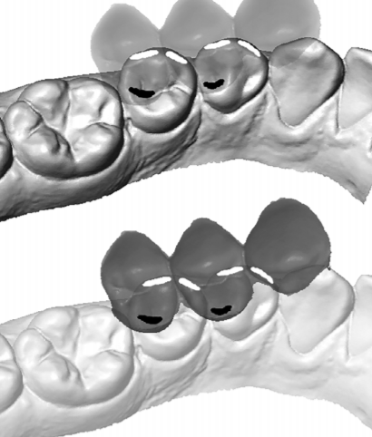
Variation of 3D outer and inner crown morphology in modern human mandibular premolars
This study explores the outer and inner crown of lower third and fourth premolars (P3, P4) by analyzing the morphological variation among diverse modern human groups.
We studied three‐dimensional models of the outer enamel surface and the enamel–dentine junction (EDJ) from μCT datasets of 77 recent humans using both an assessment of seven nonmetric traits and a standard geometric morphometric (GM) analysis. For the latter, the dental crown was represented by ... Read more
Viktoria A. Krenn, Cinzia Fornai, Lisa Wurm, Fred L. Bookstein, Martin Haeusler, Gerhard W. Weber
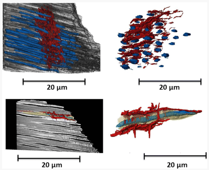
Micron-scale crack propagation in laser-irradiated enamel and dentine studied with nano-CT
The aim of this study was to see the effect of Er:YAG laser irradiation in dentine and compare this with its effect in enamel. The mechanism of crack propagation in dentine was emphasised and its clinical implications were discussed. A possible mechanism is that laser radiation is transmitted down the dentinal tubules causing micro-cracks to form in the dentinal tubule walls that tend to be limited to this region. Crack might be a source of fracture as it represents a weak point and subsequen... Read more
Abtesam Aljdaimi, Hugh Devlin, Mark Dickinson, Timothy Burnett, Thomas J. A. Slater
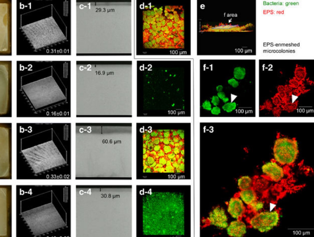
Acidic microenvironments created by bacterial clusters thriving in a polysaccharide matrix could be behind localized tooth decay. Jin Xiao of the University of Rochester Medical Center and Geelsu Hwang of the University of Pennsylvania with colleagues in the US mapped acidity changes across tooth enamel caused by the microstructure of dental plaque: a film of bacteria and the polysaccharide matrix they secrete. Using fluorescence microscopy, they studied the 3D architecture of plaque that for... Read more
Jin Xiao, Anderson T Hara, Dongyeop Kim, Domenick T Zero, Hyun Koo et al.